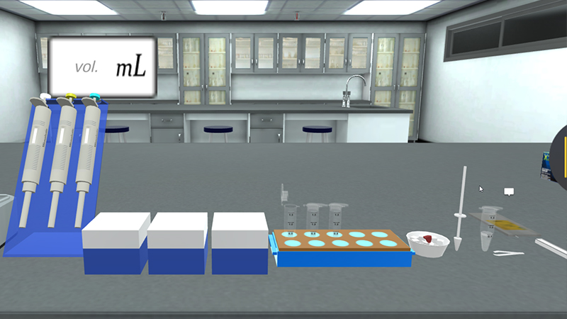
Gel Electrophoresis is a procedure used in molecular biology to separate and identify molecules (such as DNA and RNA) by size. The separation of these molecules is achieved by placing them in a gel made up of small pores and setting an electric field across the gel.
The molecules will move based on their inherent electric charge (i.e., negatively charged molecules move away from the negative pole) and smaller molecules will move faster than larger molecules; thus, a size separation is achieved within the pool of molecules running through the gel. The gel works in a similar manner to a sieve separating particles by size; the electrophoresis works to move the particles, using their inherent electric charge, through the sieve.
Agarose is isolated from the seaweed genera Gelidium and Gracilaria. It’s mixed with a buffer and heated in a microwave, then left to cool down before pouring in the cast. A comb is added at a specific site to form the wells required for sample upload. Then gel is left to solidify.
The concentration of gel = weight of agarose/volume of buffer (g/ml). For a standard agarose gel electrophoresis procedure, 0.8% gel gives good separation of large 5–10kb DNA fragments, while 2% gel gives good resolution for small 0.2-1 Kb DNA fragments.
During gelation, agarose polymers associate non-covalently and form a network whose pore sizes determine a gel's molecular sieving properties.
The phosphate in the backbone of DNA is negatively charged, therefore DNA fragments will migrate to the positively charged anode.
DNA has a uniform mass/charge ratio, therefore DNA molecules are separated by size within an agarose gel in a pattern such that the distance traveled is inversely proportional to the log of its molecular weight.
The rate of migration of a DNA molecule through a gel is determined by the following:
- Size of DNA band (the heavier, the slower).
- Agarose gel concentration (usually 0.8%).
- DNA conformation (linear/plasmid/etc..).
- Voltage.
- Electrophoresis buffer.
- Ethidium bromide: EtBr is positively charged, thus; reduces the DNA migration rate by 15%. Other stains for DNA in agarose gels include SYBR Gold, SYBR green, Crystal Violet and Methyl Blue.
UV light activates electrons in the aromatic ring of ethidium bromide releasing light as electrons return to ground state. EtBr intercalates itself in the DNA molecule in a concentration dependent manner. So higher intensity means higher amount of DNA.