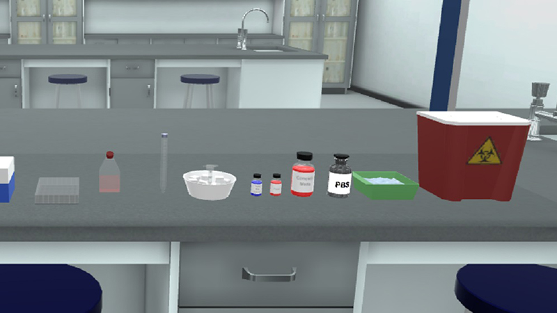
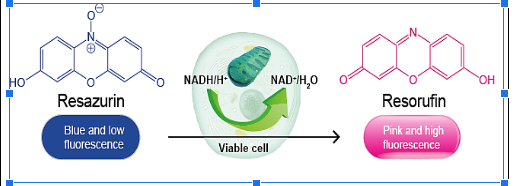
In Vitro Cell Viability by the Alamar Blue Assay

Biology | Toxicology | Biochemistry | Pharmacology

Start Now

As Featured In

Millions of Users. Better Learning. Lower Costs

icon

2.5M+

Active Users Worldwide

icon

80%

Improved Learning Retention

icon

60%

Reduction in Laboratory Costs

General Aim

  • Alamar blue assay experiment aims at describing the viability measurements for cell cultures in a 96-well tissue culture plate using Alamar Blue (Resazurin) after exposure to a geometric concentration of different nanoparticles. 
  • The alamar assay can be modified to accommodate larger plates but the 96-well plate format is the most cost-effective.

Method

In Vitro Colorimetric Analysis of Cell Viability by Alamar Blue Assay

Learning Objectives (ILOs)

  • By the end of alamar blue test experiment, students will:

  • Successfully handle the required instruments and consumables needed in the alamar blue assay experiment.
  • Check the confluence and count cells under the microscope.
  • Dilute the cells to a specific count suitable for seeding in the 96-well plate.
  • Calculate the concentration of tested chemicals and prepare the calculated doses in the cell culture medium through alamar blue assay calculation.
  • Aspirate the old medium and add the new medium containing the tested chemicals in the appropriate wells.
  • Add the Alamar Blue solution to cells and read the results using the microplate reader after incubation of cells.
  • Read the results of resorufin fluorescence and calculate the viability percent for cells exposed to different doses of tested chemical(s).

Theoretical Background / Context

  • Cytotoxicity is the quality of being toxic to cells. Cytotoxicity assays are widely used by the pharmaceutical industry to screen for cytotoxicity in compound libraries. 
  • Researchers, as in Nanotechnology, can either look for cytotoxic nano-based materials, if they are interested in developing a nanomedicine that targets rapidly dividing cancer cells, for instance; or they can screen "hits" from initial high-throughput nanoparticle screens for unwanted cytotoxic effects before investing in their development as a nanomedicine through cell cytotoxicity assay. 
  • Assessing cell membrane integrity is one of the most common ways to measure cell viability and cytotoxic effects. 
  • Compounds that have cytotoxic effects often compromise cell membrane integrity. 
  • Vital dyes, such as trypan blue or propidium iodide are normally excluded from the inside of healthy cells; however, if the cell membrane has been compromised, they freely cross the membrane and stain intracellular components. 
  • Alternatively, membrane integrity can be assessed by monitoring the passage of substances that are normally sequestered inside cells to the outside. 
  • Protease biomarkers have been identified that allow researchers to measure relative numbers of live and dead cells within the same cell population. 
  • The live-cell protease is only active in cells that have a healthy cell membrane and loses activity once the cell is compromised and the protease is exposed to the external environment. 
  • The dead-cell protease cannot cross the cell membrane, and can only be measured in culture media after cells have lost their membrane integrity. 
  • Cytotoxicity can also be monitored by measuring the reducing potential of the cells using a colourimetric reaction through colorimetric cell viability assay, or using ATP content as a marker of viability. 
  • Such ATP-based assays include bioluminescent assays in which ATP is the limiting reagent for the luciferase reaction. 
  • A label-free approach to follow the cytotoxic response of adherent animal cells in real-time provides the kinetics of the cytotoxic response rather than just a snapshot like many colourimetric endpoint assays.

Alamar Blue Assay Principle of Work

  • Resazurin is cell-permeable and virtually nonfluorescent in the resazurin cell viability assay.
  • Upon entering cells, resazurin is reduced to resorufin through the activity of cellular redox enzymes by accepting electrons from NADPH, FADH2, FMNH2, NADH, and the cytochromes (as shown in the figure) following the alamar blue assay protocol.


  • This redox reaction is accompanied by a shift in colour from indigo blue to a bright fluorescent red, which diffuses out of the cells into the culture medium in the cell membrane virtual lab, where it can be easily measured by colourimetry or fluorometry at 590 nm. 
  • Viable cells continuously convert resazurin to resorufin, thereby generating a quantitative measure of cell viability.

Trusted by Leading Universities & Institutions


What Professors Are Saying

Start the Way That Works Best for Your Institution

Start Free Trial
Schedule Personalized Demo
Get Pricing Details

Globally Recognized for Transforming STEM Education

Discover More Virtual Labs to Power Your Curriculum

Tailored Licensing to Fit Your Course, Faculty, or Whole Institution