Antibiotic Sensitivity Virtual Lab | Disc Diffusion Method
Biology | Molecular Biology | Biochemistry | Genetics | Microbiology


2.5M+
Active Users Worldwide

80%
Improved Learning Retention

60%
Reduction in Laboratory Costs
The antibiotic sensitivity virtual lab test determines the susceptibility of a microbial species against different antibiotic agents.
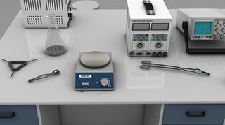
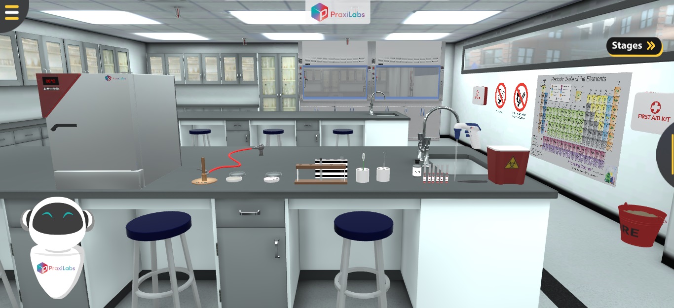
Part 1: Sterilize the bacteriological loop 1- Sterilize the bacteriological loop by holding it above the flame till it becomes red and hot. 2- Let the sterile loop cool down by holding it still. N.B. Do not wave it around to cool it or blow on it. Part 2: Antibiotic sensitivity test (Disc diffusion method) Using a sterile wire loop, pick 3–5 well-isolated colonies of similar appearance to the test organism. Then, emulsify these colonies in 4 ml of sterile physiological saline (or nutrient broth). In a good light match the turbidity of the suspension to the turbidity standard (0.5 McFarland test standard). N.B. When comparing turbidities, it is easier to view against a printed card or sheet of paper. Using a sterile swab, insert it in the test suspension. Remove excess fluid by pressing and rotating the swab against the side of the tube above the level of the suspension. Streak the swab evenly over the surface of the plate of Mueller Hinton agar in three directions, rotating the plate approximately 60o to ensure even distribution. Cover the petri dish. Allow 3–5 minutes (no longer than 15 minutes) for the surface of the agar to dry. Using sterile forceps, or a multidisc dispenser, place the appropriate, evenly distributed on the inoculated plate. You may use a template as shown in the figure will help to ensure the discs are correctly placed. N.B. The discs should be about 15 mm from the edge of the plate and no closer than about 25 mm from disc to disc. No more than 6 discs should be applied (90 mm diameter petri dish). N.B. Each disc should be lightly pressed down to ensure its contact with the agar. It should not be moved once in place. 10- Within 30 minutes of applying the discs, invert the plate and incubate it aerobically at 35o C for 16–18 h in this antibiotic sensitivity virtual lab procedure. 11- After overnight incubation, using a ruler on the underside of the plate measure the diameter of each zone of inhibition in mm. N.B. The endpoint of inhibition is where growth starts. Part 3: Interpretation of zone sizes Using the Interpretative Chart, interpret the zones sizes of each antimicrobial, reporting the organism as ‘Resistant’, ‘Intermediate/Moderately susceptible’, ‘Susceptible’.
To utilize specific monitoring techniques to evaluate the susceptibility of a microbe to different antibiotics in antibiotic sensitivity lab settings.
pH
Moisture
Amount of organism


I love the idea of virtual labs. It's gonna be something that takes our R&D and work in labs to another level. And I look forward to seeing what PraxiLabs can do with it.
Michelle Anderson, Head of Innovation
IE University - Spain
Although there are now several vendors offering virtual reality software for physics labs, there is only one that offers a realistic, I feel like I’m in a real lab solution: PraxiLabs.
Dr. William H. Miner, Jr., Professor of Physics
Palm Beach State College, Boca Raton, FL
PraxiLabs offered my students a chance to actively engage with the material. Instead of watching videos on a topic, they could virtually complete labs and realize the practical applications of class topics. This is a quality alternative to in-person labs.
Crys Wright, Teaching Assistant
Texas A&M University, USA
With the onset of the COVID-19 pandemic, we found ourselves in a situation that forced us to act quickly to find the best solution available to provide our students with a quality molecular genetics laboratory experience.
Korri Thorlacius, B.Sc., Biology Lab. Instructor
Biology Department - Kwantlen Polytechnic University