Agarose Gel Electrophoresis of Plasmid DNA - Cloning
Biology | Biochemistry | Genetics | Microbiology


2.5M+
Active Users Worldwide

80%
Improved Learning Retention

60%
Reduction in Laboratory Costs
To visualize the DNA fragments of the isolated restricted plasmids through agarose gel electrophoresis of plasmid DNA to be able to choose one for sequencing.
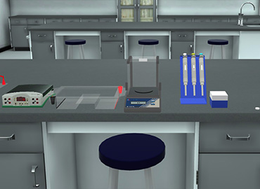
Agarose Gel Electrophoresis.
To prepare the agarose gel properly according to the agarose gel electrophoresis of plasmid dna protocol.
The purified plasmids from unit 4 were digested using restriction enzyme digest.
In this unit, you will run the restricted purified plasmids on agarose gel electrophoresis to be able to visualize DNA fragments and determine if you were successful in obtaining a recombinant DNA molecule through plasmid DNA gel electrophoresis.
Gel Electrophoresis is a procedure used in molecular biology to separate and identify molecules (such as DNA) by size.
The separation of these molecules is achieved by placing them in a gel made up of small pores and setting an electric field across the gel.
The molecules will move based on their inherent electric charge (i.e., negatively charged molecules move away from the negative pole) and smaller molecules will move faster than larger molecules; thus, a size separation is achieved within the pool of molecules running through the gel.
The gel works in a similar manner to a sieve separating particles by size.
DNA possesses negative charges. Thus, DNA moves towards the positive end (anode) when a current is applied across the gel in plasmid gel electrophoresis.
The agarose contains SYBR Safe DNA Stain which intercalates into the DNA, allowing visualization using a blue light transilluminator in the agarose gel electrophoresis of plasmid DNA.
A DNA size marker is a large piece of DNA that has been digested with one or more restriction enzymes to produce a known array of DNA fragments that range in size.
Loading a size marker in the reference lane allows you to determine the approximate sizes of the DNA fragments from your sample in the plasmid run on agarose gel.


I love the idea of virtual labs. It's gonna be something that takes our R&D and work in labs to another level. And I look forward to seeing what PraxiLabs can do with it.
Michelle Anderson, Head of Innovation
IE University - Spain
Although there are now several vendors offering virtual reality software for physics labs, there is only one that offers a realistic, I feel like I’m in a real lab solution: PraxiLabs.
Dr. William H. Miner, Jr., Professor of Physics
Palm Beach State College, Boca Raton, FL
PraxiLabs offered my students a chance to actively engage with the material. Instead of watching videos on a topic, they could virtually complete labs and realize the practical applications of class topics. This is a quality alternative to in-person labs.
Crys Wright, Teaching Assistant
Texas A&M University, USA
With the onset of the COVID-19 pandemic, we found ourselves in a situation that forced us to act quickly to find the best solution available to provide our students with a quality molecular genetics laboratory experience.
Korri Thorlacius, B.Sc., Biology Lab. Instructor
Biology Department - Kwantlen Polytechnic University