Home | Simulations
Virus Simulator - Cultivation and Preparation of the Virus in Chick Embryo

Biology | Biochemistry | Genetics | Microbiology

As Featured In

Virus Simulator - Cultivation and Preparation of the Virus in Chick Embryo

General Aim of Virus Simulator

The primary purpose of virus cultivation is to isolate and identify viruses in clinical samples; to do research on viral structure, replication, genetics and effects on host cell; to prepare viruses for vaccine production following a strict virus preparation protocol.

Method

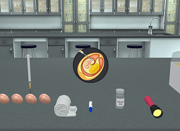
Cultivation and preparation of the virus in chick embryo: Fertile eggs are cleaned, dried, and rubbed with alcohol. 2. The date is recorded on each egg. 3. Eggs are incubated at 37 °C for 7-12 days. 4. Eggs are gently rocked daily. 5. Be sure of the growth of the embryo (beating heart) by examining the egg against a strong light in a dark chamber as shown in the virus simulator model. 6. Two points are marked on each egg under transillumination, one of them delimiting the air sac and the other opposite the semitransparent area in the neighborhood of the embryo. 7. The inoculum is introduced through an opening made in the shell using the egg shell puncher. 8. There are five main routes of virus inoculation using a syringe containing the virus: a. Into the chorioallantoic membrane. b. Into the amniotic cavity. c. Into the allantoic cavity. d. Into the yolk sac. e. Into the embryo itself. 9. After inoculation, the eggs are reincubated at 37oC for several days and examined daily in a virus virtual lab setting. 10. Virus growth is recognized by the development of: Plaques on the chorioallantoic membrane. Haemagglutinin in the amniotic or allantoic cavities. Death of the embryo (heart beats stop): just this will be shown in the experiment using a virus simulator.

Learning Objectives (ILOs)

  • Become proficient at performing the viral cultivation consistently and accurately.

  • Students will learn the essential concepts of virology in a virus simulation lab, using a virus simulator for practical understanding.

  • Learn how to use a virus simulator.

Virus Cultivation In EggTheory

  • Isolation of viruses is always considered as a gold standard for establishing viral etiology of a disease. 

Most of the viruses can be cultivated in:

  1. Experimental animals
  2. Embryonated eggs or
  3. Tissue culture
  • Experimental animals are rarely used for the cultivation of viruses but play an essential role in the study of pathogenesis of viral infections and that of viral oncogenesis.
  • Embryonated chick egg was used first for the cultivation of viruses by Goodpasture in 1931.
  • The method further developed by Burnet was used for cultivation of viruses in different sites of the embryonated egg.
  • Usually, 8–11 days’ old chick eggs are used for culture of viruses.
  • The viruses are isolated in different sites of the egg, such as yolk sac, amniotic cavity, allantoic cavity, and chorioallantoic membrane (CAM).
  • Many of these viruses cause well-defined and characteristic foci, providing a method for identification, quantification, or assessing virus pathogenicity.
  • The embryonated egg is also used for growing higher titre stocks of some viruses in research laboratories and for vaccine production.
  • Yolk sac: Yolk sac inoculation is used for cultivation of Japanese encephalitis, Saint Louis encephalitis, and West Nile virus. 
  • It is also used for growth of chlamydia and rickettsia.
  • Amniotic cavity: Inoculation in the amniotic cavity is used mainly for primary isolation of influenza virus.

Allantoic Cavity: 

  • Inoculation in the allantoic cavity is used for serial passages and for obtaining large quantities of virus, such as influenza virus, yellow fever (17D strain), and rabies (Flury strain) viruses for preparation of vaccines. 
  • For production of rabies virus, duck eggs were used due to their bigger size than that of a hen’s egg. 
  • This helped in production of large quantities of rabies virus, which are used for cultivation of viruses in microbiology and preparation of the inactivated non-neural rabies vaccine.

Chorioallantoic membrane: 

  • Inoculation of some viruses on the chorioallantoic membrane produced visible lesions known as pocks. 
  • Each infectious virus particle produces one pock. 
  • The pox viruses, such as variola or vaccinia, are identified by demonstration of typical pocks on the chorioallantoic membrane inoculated with the pox virus. 
  • Nowadays, in a virology laboratory, chick embryo inoculation has been replaced by cell cultures for routine isolation of viruses.

Principle Work of Virus Simulator

  • Viruses are obligate intracellular parasites so they depend on the host for their survival. 
  • They cannot be grown in non-living culture media or on agar plates alone; they must require living cells to support their replication.
  • Modern studies on virus-host interactions can benefit from a virus simulator to model viral

The Primary Purpose of Virus Cultivation Is:

  • To isolate and identify viruses in clinical samples.
  • To do research on viral structure, replication, genetics, and effects on host cell.
  • To prepare viruses for vaccine production.

PraxiLabs is Recognized Worldwide

Customers Love PraxiLabs

“With the onset of the COVID-19 pandemic, we found ourselves in a situation that forced us to act quickly to find the best solution available to provide our students with a quality molecular genetics laboratory experience.”

Korri Thorlacius, B.Sc.
Biology Laboratory Instructor
Biology Department
Kwantlen Polytechnic University

'' Although there are now several vendors offering virtual reality software for physics labs, there is only one that offers a realistic, I feel like I’m in a real lab, solution: PraxiLabs.''

Dr.‌ ‌William‌ ‌H.‌ ‌Miner,‌ ‌Jr.‌ ‌
Professor‌ ‌of‌ ‌Physics‌ ‌
Palm‌ ‌Beach‌ ‌State‌ ‌College‌ ‌
Boca‌ ‌Raton,‌ ‌FL‌

" PraxiLabs offered my students a chance to actively engage with the material. Instead of watching videos on a topic, they could virtually complete labs and realize the practical applications of class topics. This is a quality alternative to in-person labs."

Crys Wright
Teaching Assistant
Texas A&M University, USA

"Great user experience and impressive interaction, I am very pleased to have tried the simulations and will continue to do so."

Dr. Khaled M Goher
Lecturer in Biomedical Engineering
Aston University, UK

The #1 Science Virtual Labs used by Educational Institutions

Explore More Interactive 3D Virtual Simulations

Designed for Safety and Engagement

Find out how PraxiLabs keeps students engaged and improves learning outcomes