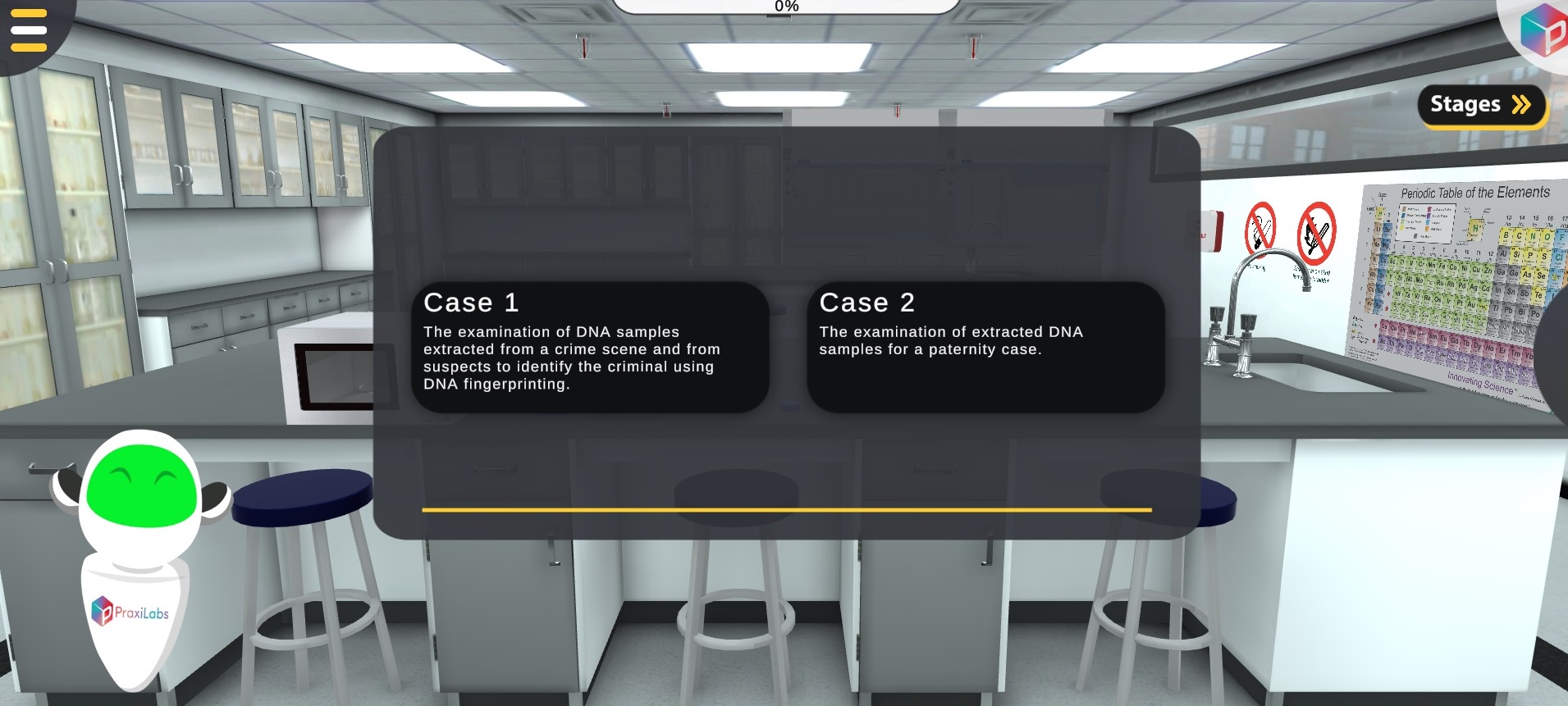
DNA Fingerprinting using Gel Electrophoresis Lab | PraxiLabs

DNA fingerprinting using Gel Electrophoresis

Biology | Molecular Biology | Biochemistry | Genetics | Microbiology

As Featured In

General Aim

Scenario One: Use DNA samples extracted from a crime scene and from suspects to identify the criminal using DNA fingerprinting. In your virtual laboratory, you will use restriction enzymes to digest the isolated DNA samples at specific sites. You will run the digested DNA in agarose gel electrophoresis to be able to see the DNA bands resulting from digestion and compare those from the suspects to those extracted from the crime scene. 
Scenario Two: Examine extracted DNA samples for a paternity case.

Method

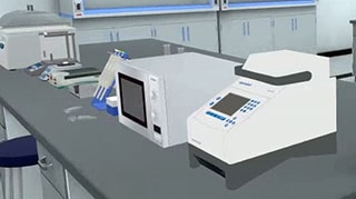
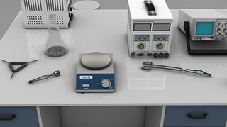
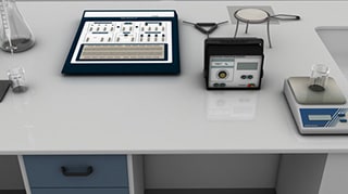
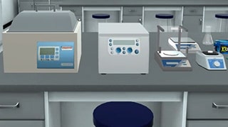
RFLP Gel Electrophoresis

Learning Objectives (ILO’s)

  • List the steps required for the preparation of an optimum agarose gel.

  • Evaluate the role and value of chemicals and reagents used in the experiment.

  • Illustrate the setup required for gel electrophoresis.

  • Enlist factors that lead to successful sample upload into the gel Visualize DNA fragments.

  • Identify and distinguish DNA molecules that have been processed by a previous method such as PCR and enzymatic digestion.

  • Interpret results on a gel UV transilluminator and solve medico-legal cases.

  • Identify the characteristics of restriction sites in a DNA sequence.

Theoretical Background/Context

Gel Electrophoresis is a procedure used in molecular biology to separate and identify molecules (such as DNA and RNA) by size. The separation of these molecules is achieved by placing them in a gel made up of small pores and setting an electric field across the gel. 
The molecules will move based on their inherent electric charge  (i.e., negatively charged molecules move away from the negative pole) and smaller molecules will move faster than larger molecules; thus, a size separation is achieved within the pool of molecules running through the gel. The gel works in a similar manner to a sieve separating particles by size;  the electrophoresis works to move the particles, using their inherent electric charge, through the sieve.  

DNA fingerprinting allows the comparison of DNA from different organisms and the identification of a particular individual. Basically, DNA is extracted and cut into fragments using restriction enzymes. The fragments form a pattern on agarose gel electrophoresis according to their length. The longer the DNA fragments are generated, the larger their molecular weight and the shorter they travel in an electrophoresis setting. This pattern looks a lot like the barcode on products in the supermarket and it resembles the individual’s DNA fingerprint.

To interpret the pattern formed, one must realize that restriction enzymes cut the DNA at specific base-pair sequences called recognition sequences. There are hundreds of restriction enzymes, each having a specific recognition sequence made of four to twelve base pairs. The lengths of the fragments generated by a restriction enzyme digest of an extracted DNA sample depend upon the number of cuts made and their locations. This depends on the number of recognition sequences of the enzyme used on the extracted DNA and their locations. Thus, everyone’s DNA is cut by restriction enzymes into distinctive different sized fragments that appear on the electrophoresis gel in the form of bands.


Principle of Work

Agarose is isolated from the seaweed genera Gelidium and Gracilaria. It’s mixed with a buffer and heated in a microwave, then left to cool down before pouring in the cast. A comb is added at a specific site to form the wells required for sample upload. Then the gel is left to solidify. 
The concentration of gel = weight of agarose/volume of buffer (g/ml). For a standard agarose gel electrophoresis, 0.8% gel gives good separation of large 5–10kb DNA fragments, while 2% gel gives a good resolution for small 0.2-1 Kb DNA fragments.
During gelation, agarose polymers associate non-covalently and form a network whose pore sizes determine a gel's molecular sieving properties. 
 
 
The phosphate in the backbone of DNA is negatively charged, therefore DNA fragments will migrate to the positively charged anode. 
DNA has a uniform mass/charge ratio, therefore DNA molecules are separated by size within an agarose gel in a pattern such that the distance traveled is inversely proportional to the log of its molecular weight. 
The rate of migration of a DNA molecule through a gel is determined by the following: 
1) size of DNA band (the heavier, the slower)
2) agarose gel concentration (usually 0.8%)
3) DNA conformation (linear/plasmid/etc..)
4) Voltage 
5) Electrophoresis buffer.
6) Ethidium bromide: EtBr is positively charged, thus; reducing the DNA migration rate by 15%. Other stains for DNA in agarose gels include SYBR Gold, SYBR green, Crystal Violet and Methyl Blue. 
 
UV light activates electrons in the aromatic ring of ethidium bromide releasing light as electrons return to the ground state. EtBr intercalates itself in the DNA molecule in a concentration-dependent manner. So higher intensity means a higher amount of DNA.


Restriction enzymes are present in bacteria. They are named according to the bacteria from which they are extracted. They protect bacteria by digesting foreign DNA at specific sequences.
Restriction enzymes such as (EcoRI) and (HindIII) are used in this experiment to Cut the extracted DNA. EcoRI recognizes the 6 bp sequence 5’ GAATTC 3’ and makes a staggered cut between the G and A creating sticky ends. HindIII recognizes the 6 bp sequence 5’ AAGCTT 3’ and makes a staggered cut between the A and A creating sticky ends. Both enzymes cut best at 37°C. 

EcoRI:
5’ G^AATTC 3’
3’ CTTAA^G 5’

HindIII:
5' A ↓AGCTT 3' 
3' TTCGA ↑A 5'

PraxiLabs is Recognized Worldwide

Customers Love PraxiLabs

“With the onset of the COVID-19 pandemic, we found ourselves in a situation that forced us to act quickly to find the best solution available to provide our students with a quality molecular genetics laboratory experience.”

Korri Thorlacius, B.Sc.
Biology Laboratory Instructor
Biology Department
Kwantlen Polytechnic University

'' Although there are now several vendors offering virtual reality software for physics labs, there is only one that offers a realistic, I feel like I’m in a real lab, solution: PraxiLabs.''

Dr.‌ ‌William‌ ‌H.‌ ‌Miner,‌ ‌Jr.‌ ‌
Professor‌ ‌of‌ ‌Physics‌ ‌
Palm‌ ‌Beach‌ ‌State‌ ‌College‌ ‌
Boca‌ ‌Raton,‌ ‌FL‌

" PraxiLabs offered my students a chance to actively engage with the material. Instead of watching videos on a topic, they could virtually complete labs and realize the practical applications of class topics. This is a quality alternative to in-person labs."

Crys Wright
Teaching Assistant
Texas A&M University, USA

"Great user experience and impressive interaction, I am very pleased to have tried the simulations and will continue to do so."

Dr. Khaled M Goher
Lecturer in Biomedical Engineering
Aston University, UK

The #1 Science Virtual Labs used by Educational Institutions

Explore More Interactive 3D Virtual Simulations

Designed for Safety and Engagement

Find out how PraxiLabs keeps students engaged and improves learning outcomes