


2.5M+
Active Users Worldwide

80%
Improved Learning Retention

60%
Reduction in Laboratory Costs
Gene expression profiling
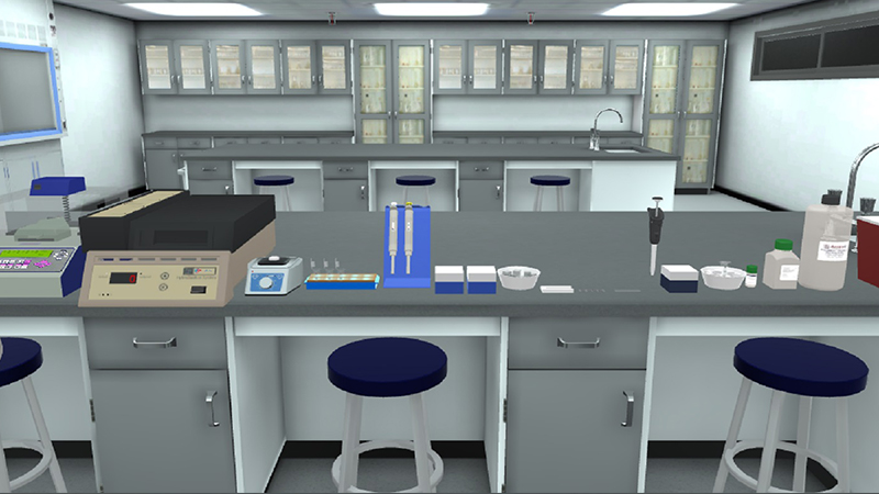
DNA Microarray Assay hybridization: whole human genome (4*44) oligo microarray and dual-color labelling kits.
By the end of DNA Microarray virtual lab, students will be able to:
Other applications of DNA microarray Experiment:
In this DNA Microarray virtual lab protocol, Two RNA samples were prepared: cancerous & healthy (reference).
A- Amplification and labeling of extracted RNA:
1) synthesizing cDNA:
Using T7 RNA polymerase by reverse transcription.
2) synthesizing cRNA:
cDNA is used in an in vitro transcription reaction to generate cRNA. This reaction is performed in the presence of labeled ribonucleotides, producing microgram quantities of labeled RNA for array hybridization.
B- Microarray hybridization:
A hybridization sample is prepared and added to the cRNA prior to array hybridization. During this stage, cRNA is fragmented and labeled. cyanine 3 (cy3-fluoresces green) is added to the ‘to-be studied’ cells and cyanine 5 (cy5-fluoresces red) is added to the reference cells.
C- Microarray slide wash prior to scan:
The scanner has a laser which causes the hybrid bonds to fluoresce


I love the idea of virtual labs. It's gonna be something that takes our R&D and work in labs to another level. And I look forward to seeing what PraxiLabs can do with it.
Michelle Anderson, Head of Innovation
IE University - Spain
Although there are now several vendors offering virtual reality software for physics labs, there is only one that offers a realistic, I feel like I’m in a real lab solution: PraxiLabs.
Dr. William H. Miner, Jr., Professor of Physics
Palm Beach State College, Boca Raton, FL
PraxiLabs offered my students a chance to actively engage with the material. Instead of watching videos on a topic, they could virtually complete labs and realize the practical applications of class topics. This is a quality alternative to in-person labs.
Crys Wright, Teaching Assistant
Texas A&M University, USA
With the onset of the COVID-19 pandemic, we found ourselves in a situation that forced us to act quickly to find the best solution available to provide our students with a quality molecular genetics laboratory experience.
Korri Thorlacius, B.Sc., Biology Lab. Instructor
Biology Department - Kwantlen Polytechnic University