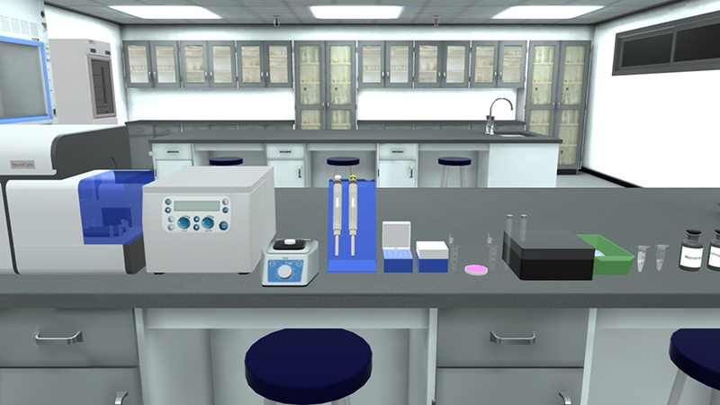
Flow cytometry is a laser-based technology that allows quantitative single cell analysis. It is used to analyze the characteristics of cells. Analysis of the Cell cycle by DNA quantification can be done using flow cytometry. The DNA in cells can be stained by DNA binding dyes e.g. Propidium iodide. These dyes bind in proportion to the amount of the DNA present in the cell.
Cells in S phase have more DNA than cells in G1 phase. Cells in G2 will have approximately twice DNA content as cells in G1. Thus; they will take up proportionately more dye. The suspension of cells is then aspirated into a flow cell. Cells, surrounded by a narrow fluid stream, pass one by one through a focused laser beam. The light is either scattered or absorbed when it strikes a cell.
Absorbed light of the appropriate wavelength is reemitted as fluorescence. This reflects the internal structure of the cell and its size and shape. Fluorescence scatter signals are detected, amplified and analyzed by a series of photodiodes and a computer system.
The end result is quantitative information about every cell analyzed. Large numbers of cells are analyzed in a short period of time (>1,000/sec). This gives the advantage of creating statistically valid information about cell populations.
The potential applications of flow cytometry include the detection and measurement of:
1-Cell cycle: Reliable assessment of cells in G0/G1 phase versus S phase, G2, or polyploidy, including analysis of cell proliferation and activation.
2-Cell viability/apoptosis.
3-Identification and characterization of distinct subsets of cells within a heterogeneous sample.
4-Protein expression.
5-Protein post translational modifications.
6-RNA, including IncRNA, miRNA, and mRNA transcripts.