


2.5M+
Active Users Worldwide

80%
Improved Learning Retention

60%
Reduction in Laboratory Costs
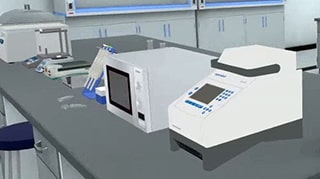
Hemagglutination used for the diagnosis of some enveloped viruses such as influenza viruses.
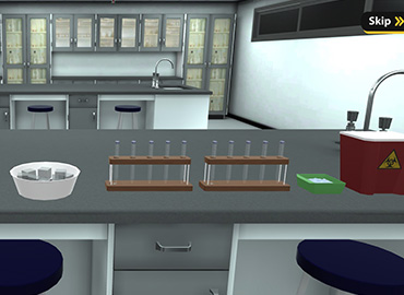
The technique of Haemagglutination Test : Haemagglutination test is used to quantify the amount of animal virus in a suspension. This is done by carrying out two-fold serial dilutions of the viral suspension in a microtitre plate or test tubes and then testing to determine an endpoint. This result can then be used to determine the amount of haemagglutinin in the suspension and is expressed as a HA titre. One HA unit in the haemagglutinin titration is the minimum amount of virus that will cause agglutination of the red blood cells. 1. Prepare 10 sterile test tubes. 2. Add 0.5 ml of normal saline to each of the test tubes. 3. Add 0.5 ml of undiluted virus to the first tube ONLY and mix gently. 4. Transfer 0.5 ml to the next tube and mix gently. 5. Repeat this procedure for the whole test tubes, discarding 0.5 ml from the last tube (8th tube). 6. This will lead to bifold dilution of the virus starting from 1:2, 1:4, 1:8, 1:16, 1:32, 1:64, 1:128 & 1:256. So the dilution in the 1st tube is 1:2 , in the 2nd tube is 1:4, in the 3rd tube is 1:8, in the 4th tube is 1:16, and so on. 7. Using a clean pipette tip, add 0.5 ml of 1% fresh RBCs suspension to each tube. 8. Two negative control test tubes (9th and 10th tubes) are prepared: one consisting of a mixture of normal saline and RBCs and the other of a mixture of non-infected allantoic fluid with the RBCs. 9. Allow the tubes to stand on ice for 45 minutes. 10. Read and record the results in each well. 11. Identify the endpoint. This will be the last well to show complete haemagglutination and contains one haemagglutinating unit.
By the end of hemagglutinin test, student will:
Then, aliquots of RBC are added to each well. The highest dilution at which clumping is observed is regarded as the HA titer of the sample. The virus titer in a sample can be estimated by multiplying the dilution fold.


I love the idea of virtual labs. It's gonna be something that takes our R&D and work in labs to another level. And I look forward to seeing what PraxiLabs can do with it.
Michelle Anderson, Head of Innovation
IE University - Spain
Although there are now several vendors offering virtual reality software for physics labs, there is only one that offers a realistic, I feel like I’m in a real lab solution: PraxiLabs.
Dr. William H. Miner, Jr., Professor of Physics
Palm Beach State College, Boca Raton, FL
PraxiLabs offered my students a chance to actively engage with the material. Instead of watching videos on a topic, they could virtually complete labs and realize the practical applications of class topics. This is a quality alternative to in-person labs.
Crys Wright, Teaching Assistant
Texas A&M University, USA
With the onset of the COVID-19 pandemic, we found ourselves in a situation that forced us to act quickly to find the best solution available to provide our students with a quality molecular genetics laboratory experience.
Korri Thorlacius, B.Sc., Biology Lab. Instructor
Biology Department - Kwantlen Polytechnic University