In Vitro Micronucleus Assay Theoretical Background / Context
- In genetics, genotoxicity describes the property of chemical agents that damages the genetic information within a cell causing mutations, which may lead to cancer.
- While genotoxicity is often confused with mutagenicity, all mutagens are genotoxic, whereas not all genotoxic substances are mutagenic. The alteration can have direct or indirect effects on the DNA: the induction of mutations mistimed event activation, and direct DNA damage leading to mutations.
The permanent, heritable changes can affect either somatic cells of the organism or germ cells to be passed onto future generations. Cells prevent expression of the genotoxic mutation by either DNA repair or apoptosis; however, the damage may not always be fixed leading to mutagenesis.
Principle of Work of Micronucleus Test In Vitro
- The cytokinesis-block micronucleus assay (CBMN) is a sensitive and simple indicator of chromosome damage, both chromosome loss and chromosome breakage, which also provides information on cell cycle progression and cytotoxicity.
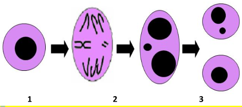
- MNs are small nuclei separated from the main nucleus and contain chromosomes or chromosome fragments, derived from mitotic spindle dysfunction or acentric fragments.
- Because they are expressed in cells that have completed nuclear division, they are ideally scored in the binucleated stage of the cell cycle.

Figure 1: Cell starts its cell cycle and replication of its genetic materials (1), under various genotoxic stresses, (Chromosomal damage occurs during replication) chromosomes are damaged during replication (2), the damaged fragments are not segregated by spindles and form micronuclei (3), and Micronuclei are separated into divided cells (4)
- The cytokinesis block (CB) method can distinguish, within a cell population, between cells that are not dividing and cells that are undergoing mitosis.
- The CBMN test is the most suitable assay due to its versatility, simplicity, and lack of effects on baseline genetic damage.
- This technique consists in adding to cell cultures cytochalasin-B (Cyt-B), an inhibitor of the mitotic spindle that prevents cytokinesis. As a consequence, cells that have completed one nuclear division are identified by their binucleated appearance.
- Micronuclei inside binucleated cells are then stained with Giemsa stain and visualized by a light microscope. Alternatively, cells could be stained by acridine orange and visualized by a fluorescent microscope.