Western Blot Virtual Lab Simulation | PraxiLabs

Western blot

Biology | Biochemistry | Proteomics


As Featured In

Western Blot: What You Need to Know (H2)

Importance of Western Blot

To detect a specific protein in a sample using western blot technique.

Western Blot Method

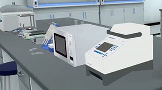
Electroblotting of proteins into nitrocellulose membrane.

Western Blotting Technique: Learning Objectives for Students

  • <p>To identify the theory behind western blot technique.

  • To fully comprehend the Western blot steps.

  • To design a complete western blot experiment.

  • To analyze the visualized protein and understand Western blotting application.</p>

Western Blot Experiment: Theoretical Background

A blot, in molecular biology, is the technique that involves the transfer of DNA, RNA and proteins from a gel to a membrane (nitrocellulose or PVDF). Proteins can be detected by western blot (immunoblot). Labeled probes or antibodies can bind blotted proteins and reveal them, making it easier to study them.

The Difference between Elisa and Western Blot

The ELISA test uses enzymes or antibodies attached to a solid surface to create the test surface. A sample is then added to the test surface. Antibodies or enzymes that react or attach to proteins indicate a positive result. The Western blot test is performed after gel-electrophoresis. The separated proteins are transferred (or blotted) onto nitrocellulose or nylon membranes, which are then identified by specific antibodies that are tagged by a secondary protein. It detects viral antigens (proteins usually on the surface of viruses) using antibodies against those proteins. A positive Western blot indicates viral antigen presence.

Western Blot Experiment Principle of Work

Western blot is a commonly used technique designed to study a specific protein in a sample containing many other proteins. Proteins in the sample are extracted then separated by gel electrophoresis (SDS-PAGE) according to their molecular weight. Then, the proteins are electrophoretically transferred (blotted) to a more durable surface (e.g. nitrocellulose or PVDF membrane). Finally, a specific antibody is used to detect the protein of interest. The bound and labeled protein can be visualized giving information about its molecular weight and relative quantity.


PraxiLabs is Recognized Worldwide

Customers Love PraxiLabs

“With the onset of the COVID-19 pandemic, we found ourselves in a situation that forced us to act quickly to find the best solution available to provide our students with a quality molecular genetics laboratory experience.”

Korri Thorlacius, B.Sc.
Biology Laboratory Instructor
Biology Department
Kwantlen Polytechnic University

'' Although there are now several vendors offering virtual reality software for physics labs, there is only one that offers a realistic, I feel like I’m in a real lab, solution: PraxiLabs.''

Dr.‌ ‌William‌ ‌H.‌ ‌Miner,‌ ‌Jr.‌ ‌
Professor‌ ‌of‌ ‌Physics‌ ‌
Palm‌ ‌Beach‌ ‌State‌ ‌College‌ ‌
Boca‌ ‌Raton,‌ ‌FL‌

" PraxiLabs offered my students a chance to actively engage with the material. Instead of watching videos on a topic, they could virtually complete labs and realize the practical applications of class topics. This is a quality alternative to in-person labs."

Crys Wright
Teaching Assistant
Texas A&M University, USA

"Great user experience and impressive interaction, I am very pleased to have tried the simulations and will continue to do so."

Dr. Khaled M Goher
Lecturer in Biomedical Engineering
Aston University, UK

The #1 Science Virtual Labs used by Educational Institutions

Explore More Interactive 3D Virtual Simulations

Designed for Safety and Engagement

Find out how PraxiLabs keeps students engaged and improves learning outcomes