


2.5M+
Active Users Worldwide

80%
Improved Learning Retention

60%
Reduction in Laboratory Costs
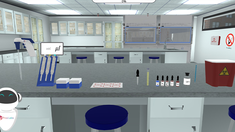
The Widal test can be conducted in two ways: Slide agglutination Widal test 1. Qualitative Slide Test 2. Quantitative Slide Test Tube agglutination Widal test Tube agglutination has more accuracy as compared to the slide agglutination technique. However, A slide Widal test is more popular among diagnostic laboratories as it gives rapid results. Part 1 Qualitative Slide Test Procedure: Bring all reagents (Widal kit) to room temperature and mix well by shaking. Add 1 drop of test serum sample (25µl) into each reaction circle labeled as O, H, AH, BH according to the given antigen solution. Add 1 drop of positive control (25µl) into the circle marked as PC. Add 1 drop of physiological saline which serves as a negative control (25µl) into the reaction circle marked as NC. Add 1 drop of Salmonella O antigen to the reaction circles PC and NC. Add antigen solutions of Salmonella typhi ‘O’, Salmonella typhi ‘H’, Salmonella paratyphi ‘AH’ and Salmonella paratyphi ‘BH’ to circles labeled as O, H, AH and BH respectively in which test samples have been added. Mix it thoroughly with the aid of separate applicator sticks and rotate the slide gently. Observe for agglutination at 1 minute (adjust a timer). Interpretation: Positive Test: Agglutination within a minute Negative Test: No agglutination Part 2 Quantitative Slide Test: This is performed for the samples which showed positive agglutination during the qualitative slide test. Procedure: Bring all reagents to room temperature and mix well. Using a pipette place 80, 40, 20, 10, and 5 ul of the test sample on circles labeled 1/20,1/40,1/80, 1/160, and 1/320 respectively. Add a drop of the antigen, which showed agglutination with the test sample in the screening (qualitative) method, to each circle. Mix the contents of each circle with the aid of separate applicator sticks and rotate the slide gently. Observe for agglutination. Interpretation: The antibody titre of the test sample is at its highest dilution which gives a visible agglutination. 80 µl corresponds to 1 in 20 dilution, 40 µl to 1 in 40, 20 µl to 1 in 80, 10 µl to 1 in 160 and 5 µl corresponds to 1 in 320 titre. Agglutination titre greater than 1:80 is considered as a significant infection and low titres indicate the absence of infection. Part 3 Quantitative Tube Test: Procedure: Bring all reagents to room temperature and mix well. Prepare 4 sets of test tubes for individual antigen. Each set contains 1-8 tubes. Add 1.9 ml of sterile physiological saline to tube no.1 of each antigen set. To tubes no.2-8 of all sets add 1 ml of physiological saline. To tubes no.1 of all sets add 0.1 ml of a test serum sample to be tested and mix well. Transfer 1 ml of the diluted serum sample from tube no.1 to tube no.2 and mix well. Transfer 1 ml of the diluted serum sample from tube no.2 to tube no.3 and mix well. Continue this serial dilution till tube no.7 in each antigen set. Discard 1.0 ml of the diluted serum from tube no.7 of each set. So, the dilutions of the serum sample from tube no.1 to 7 respectively in each antigen set are 1:20, 1:40,1:80, 1:160, 1: 320, 1:640 and 1: 1280. Tube no.8 is negative control with sterile saline only. To one set i.e. from tube no.1- 8 add 50 µl of Salmonella typhi ‘O’ antigen. In the second set i.e. from tubes no.1- 8 add 50 µl of Salmonella typhi ‘H’ antigen. In the third set, add Salmonella paratyphi ‘AH’ to all tubes from 1-8. In the fourth set, add Salmonella paratyphi ‘BH’ to all tubes from 1-8. Mix well, cover, and incubate these tubes overnight at 370 Celsius (approximately 18 hours). After incubation, dislodge the sediment and observe for agglutination. Interpretation: The antibody titre of the test sample is at its highest dilution which gives a visible agglutination. Agglutinin titre greater than 1:80 is considered as a significant infection and low titres indicate the absence of infection.


I love the idea of virtual labs. It's gonna be something that takes our R&D and work in labs to another level. And I look forward to seeing what PraxiLabs can do with it.
Michelle Anderson, Head of Innovation
IE University - Spain
Although there are now several vendors offering virtual reality software for physics labs, there is only one that offers a realistic, I feel like I’m in a real lab solution: PraxiLabs.
Dr. William H. Miner, Jr., Professor of Physics
Palm Beach State College, Boca Raton, FL
PraxiLabs offered my students a chance to actively engage with the material. Instead of watching videos on a topic, they could virtually complete labs and realize the practical applications of class topics. This is a quality alternative to in-person labs.
Crys Wright, Teaching Assistant
Texas A&M University, USA
With the onset of the COVID-19 pandemic, we found ourselves in a situation that forced us to act quickly to find the best solution available to provide our students with a quality molecular genetics laboratory experience.
Korri Thorlacius, B.Sc., Biology Lab. Instructor
Biology Department - Kwantlen Polytechnic University