


2.5M+
Active Users Worldwide

80%
Improved Learning Retention

60%
Reduction in Laboratory Costs
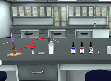
⦁ Place a slide with a heat fixed smear on a staining tray. ⦁ Gently flood the smear with concentrated carbol fuchsin. ⦁ Heat the stain until vapour just begins to rise. N.B. Do not overheat ⦁ Heating the stain: Great care must be taken when heating the carbol fuchsin. Only a small flame should be applied under the slides using an ignited swab previously dampened with a few drops of acid alcohol or 70% ethanol or methanol. ⦁ Do not use a large ethanol-soaked swab because this is a fire risk. ⦁ Allow the heated stain to remain on the slide for 5 minutes. ⦁ Tilt the slide slightly and gently rinse with tap water (or distilled water using a wash bottle if the tap water is not clean) N.B. While washing the slide after staining, do not let the water stream fall directly on the smear. This may disrupt the smear. Let the stream of water flow slowly along the surface, such that only the stain is flooded, and the smear is intact. ⦁ The smear will appear as a red circle on the slide. ⦁ Decolorize using 20% H2SO4 (or 3% HCL). Add the acid and leave it for 1-2 minutes. ⦁ Repeat this step until the smear appears pink in color. Caution: Acid is flammable; therefore use it with care well away from an open flame. ⦁ Immediately rinse with water. ⦁ Flood the smear with methylene blue dye and leave it for 1-2 minutes. ⦁ Tilt the slide slightly and gently rinse with tap water or distilled water using a wash bottle. ⦁ The smear will appear as a blue circle on the slide. ⦁ Blot dry the slide with bibulous paper. ⦁ View the smear using a light-microscope under oil-immersion lens (100x).
To become proficient at performing the Ziehl Neelsen stain consistently and accurately following the ZN staining procedure.


I love the idea of virtual labs. It's gonna be something that takes our R&D and work in labs to another level. And I look forward to seeing what PraxiLabs can do with it.
Michelle Anderson, Head of Innovation
IE University - Spain
Although there are now several vendors offering virtual reality software for physics labs, there is only one that offers a realistic, I feel like I’m in a real lab solution: PraxiLabs.
Dr. William H. Miner, Jr., Professor of Physics
Palm Beach State College, Boca Raton, FL
PraxiLabs offered my students a chance to actively engage with the material. Instead of watching videos on a topic, they could virtually complete labs and realize the practical applications of class topics. This is a quality alternative to in-person labs.
Crys Wright, Teaching Assistant
Texas A&M University, USA
With the onset of the COVID-19 pandemic, we found ourselves in a situation that forced us to act quickly to find the best solution available to provide our students with a quality molecular genetics laboratory experience.
Korri Thorlacius, B.Sc., Biology Lab. Instructor
Biology Department - Kwantlen Polytechnic University