Last Updated on September 17, 2025 by Muhamed Elmesery
Pharmacology plays a crucial and vital role in modern medicine. From ancient remedies to modern drugs, pharmacology has evolved significantly over the centuries. It studies how drugs interact with different living organisms and how they can be used to treat and prevent diseases for a healthier life. In this article, we will take a closer look at the science of pharmacology, its definition, branches, history, and importance. We will also show you some examples of pharmacology experiments that are introduced by PraxiLabs virtual labs.
Table of Contents
Pharmacology Definition
In simple words, pharmacology is the science of drugs. Also, we can define pharmacology as the science that studies the chemistry, origin, effects, properties, mechanisms, toxicity, and different uses of drugs. It is the study of chemicals and living organisms’ interactions and the effect of chemicals on the biochemical function whether it is normal or abnormal.
What is Pharmacology?
Pharmacology is considered as a branch of biology and medicine that deals with drug action.
Note that a drug is any molecule (natural, artificial, or endogenous from within the body) that exerts physiological or biochemical effects on the cells, tissues, organs, or organisms.
Types of Pharmacology
There are 2 main Branches of Pharmacology:
Pharmacokinetics
“Pharmacokinetics” word is derived from 2 words, Pharmacon which means drug and kinetics which means putting in motion. Pharmacokinetics refers to what our bodies do to the drugs we consumed like distribution, absorption, metabolism and extraction processes.
Pharmacodynamics
Pharmacodynamics is the branch of Pharmacology that deals with the drugs mechanism of action. It studies the effects of a drug on biological systems ex: our bodies, microorganisms, and parasites. Pharmacodynamics refers to what drugs do to the body.
Other types of pharmacology are:
Clinical Pharmacology
Clinical pharmacology is the scientific study of medication in man. It includes mainly pharmacodynamics and pharmacokinetic investigations in both healthy and diseased humans. It also deals with detecting the effective, safe, and economical use of drugs in patients.
The main objectives of clinical pharmacology are minimizing the adverse effects of medications and maximizing the effect of drugs, ensuring and promoting the safety of prescriptions.
The other branches of pharmacology are Therapeutics/ Toxicology / Pharmacogenetics/ Pharmacy/Chemotherapy/ Clinical Pharmacology / Pharmacognosy/ Pharmacoeconomics/ Pharmacoepidemiology/ Animal Pharmacology/ Pharmacoeconomics/ Posology/ Comparative Pharmacology.
PraxiLabs’ pharmacology virtual labs, students can practice the same experiment an unlimited number of times, with 100% supervision & 0% risk!
History of Pharmacology
The following video describes the history of pharmacology
The Importance of Pharmacology
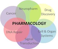
Pharmacology helps people to improve their life and live healthier for longer. We can find the importance of pharmacology everywhere. It has a vital role in:
- Discovering new drugs to help in fighting diseases and recovery.
- Understanding why some drugs can cause addiction and why drugs work better for some people than others.
- The highly sensitive drugs with fewer side effects led to reducing infectious diseases and types of cancer.
- Make sure that drugs are safe for consumption.
- Developing medicines to tackle new diseases
- Identifying and discovering new treatments.
- Researching to ensure that drugs are safe and effective for everyone.
- Exploring and developing how we can best use the medicines we already have.
- Tackling and studying important topics like aging and antibiotic resistance.
- Helping to ensure that everyone is prescribed the right drugs for them.
Pharmacology Experiments Examples Introduced by PraxiLabs
PraxiLabs provides a vast and exceptionally diverse collection of important pharmacology experiments in our virtual biology labs with awesome features to improve student’s learning experiences and outcomes.
Let’s take a look at some of PraxiLabs pharmacology virtual lab simulations!
Adherent Cell Culturing using Mammalian Cell Lines
Learn how to culture, propagate and sub-culture the cells. Discover also how cells are frozen in liquid nitrogen for the process of long-term storage.
By the end of the experiment, students will be able to:
- Apply the Aseptic Techniques of cell culturing.
- Handle the required instruments that are needed in the cell culturing and sub-culturing successfully.
- Prepare the cells needed for all subsequent In Vitro Genotoxicity and Cytotoxicity assays.
- Understand the general aim, principle, and protocol of cell culturing using mammalian cell lines experiment.
- Work and apply the general safety guidelines for Good Laboratory Practice (GLP).
- Check and count the harvest cells microscopically.
- Freeze cells in liquid nitrogen for long-term storage.
- Thaw cells from Liquid Nitrogen and then seed them in cell culture flasks.
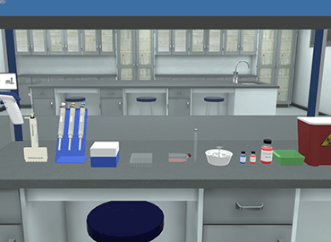
In Vitro Viability Assay Using Tetrazolium Salt XTT
In this experiment, students will test the viability of cultured cells after exposure to the geometric concentration of different nanoparticles.
By the end of the experiments students will be able to:
- Handle the required instruments which are needed in the experiment successfully.
- Check the count of cells and confluence by using the microscope.
- Understand XTT viability assay protocol, procedure, and principle of work.
- Dilute the cells to a specific count which is suitable for the process of seeding in the 96-well plate.
- Calculate the tested chemicals concentration and then prepare the required doses in the cell culture media.
- Add the solution of XTT assay to the cells and by using the microplate reader, you can read the XTT viability assay results.
- Read the results of the XTT viability assay experiment
- Do the XTT viability assay calculation: calculate the viability percent of cells exposed to different doses of tested chemicals.
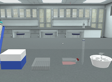
In Vitro Cell Viability by the Lactate Dehydrogenase Assay (LDH)
Learn how to determine the activity of the cytoplasmic enzyme lactate dehydrogenase (LDH) released by damaged cells after exposure to the geometric concentration of different nanoparticles.
By the end of lactate dehydrogenase assay experiment, students will be able to:
- Add the LDH assay substrate solution to cells and then by using the microplate reader, they will read the results after cell incubation.
- Prepare the LDH standard solution (in the required concentrations) in cell culture medium, then draw the standard curve.
- Read the results of LDH and calculate the percent of cytotoxicity for cells that are exposed to different doses of tested chemicals.
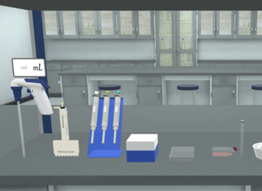
In Vitro Cell Viability by the Alamar Blue Assay
Discover how to describe the measurements of viability for cell cultures in a 96-well tissue culture plate by using Resazurin (Alamar Blue) after exposure to geometric concentration of different nanoparticles.
In this experiment, students will:
- Learn alamar blue assay procedure by adding the solution of alamar blue to the cells and after incubation of cells they caan read the results using the microplate reader.
- Understand alamar blue assay principle and steps.
- Read the results of Resorufin and calculate the percent of viability for cells which exposed to different doses of tested materials.
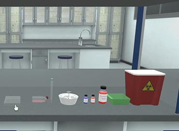
In Vitro Histone H2AX Phosphorylation Assay
In this experiment, you will find all you need to understand how to detect DNA damage and phosphorylated histone H2AX. Also how to visualize the response of protein recruitment to DNA damage sites.
By the end of the experiment, students will be able to:
- Handle the required instruments which are needed in the experiment successfully.
- Learn the right technique for adding the substrate solution of H2AX Phosphorylation assay to cells and by using the fluorescent microscope (after incubation of cells), they can read the results.
- Understand in vitro phosphorylation assay protocol and principle.
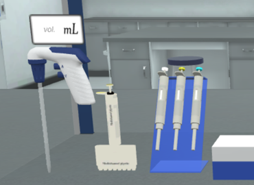
In Vitro 8-Hydroxydeoxy Guanosine (8-OHdG) Assay
Understand Detection 8-ohdg assay experiment and how to visualize the response of protein recruitment to DNA damage sites.
By the end of the experiment, students will be able to:
- Understand the principle and protocol of In Vitro 8-Hydroxydeoxy Guanosine (8-OHdG) Assay experiment.
- Add the blocking solution, first and secondary antibodies to cells and then by using the fluorescent microscope, they can read the result after incubation of cells.
Try 8-ohdg assay with Praxilabs 3D science experiments simulations now!
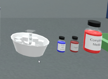
In Vitro Bromodeoxyuridin (BrdU) Assay
This experiment aims at visualizing the incorporation of Bromodeoxyuridine (BrdU) as a thymidine analog into nuclear DNA for detecting DNA synthesis in vitro by using a fluorescent microscope and antibody probes.
By the end of the experiment, students will be able to:
- Understand the principle, procedure, and protocol of Bromodeoxyuridine (BrdU) Assay.
- Add the BrdU assay substrate solution to cells and after incubation of cells with first anti-BrdU and secondary antibodies, they can read the results by using the fluorescent microscope.
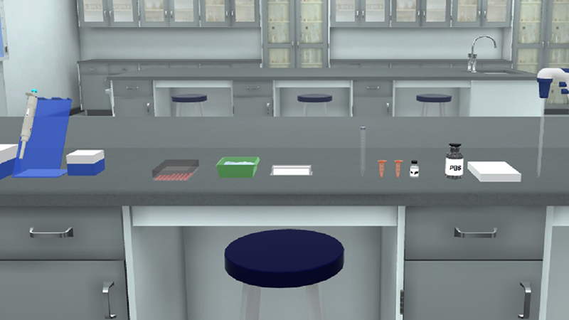
Annexin V Assay
Learn how to determine and quantify necrotic and apoptotic cells through Annexin-V binding and Propidium Iodide (PI) uptake by using a fluorescent microscope.
By the end of annexin V assay experiment, students will be able to:
- Treat cells with nanoparticles or cytotoxic agents and then observe using the microscope.
- Harvest the cells with the binding buffer of annexin (Annexin-V / Propidium Iodide buffer).
- Analyze both of cells (y using the fluorescent microscope) and resulted data.
- Represent and interpret the resulting data graphically using dot plots.
- Understand the principle and procedure of annexin V assay.
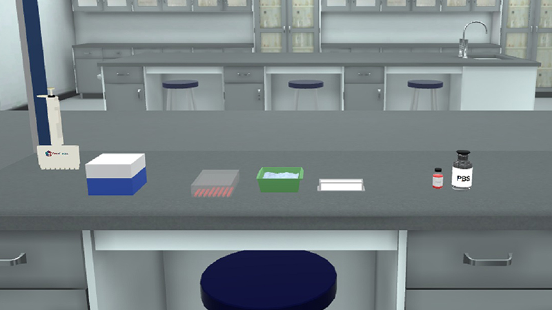
In Vitro Mammalian Cells COMET Assay
The in vitro mammalian Comet assay which is also known as the single-cell gel electrophoresis assay or (SCGE) assay is a rapid, simple, versatile, and sensitive tool that used to detect primary DNA damage at an early stage in a 96-well tissue culture plate after exposure to the geometric concentration of different nanoparticles.
By the end of the experiment, students will be able to:
- Understand the principle and procedure used in the comet assay experiment.
- Understand and follow the protocol step by step to achieve the required results.
- Read the results of cells that are exposed to different doses of tested chemicals using the COMET software and fluorescent microscope.
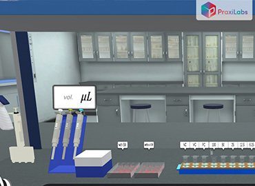
In Vitro Cytokinesis-Block Micronucleus Assay
Understand how to detect the damage of chromosomes (both chromosome loss and breakage) and how to evaluate the induction of genotoxic effects of nanomaterials and nanoparticles by using the light microscope.
By the end of Cytokinesis-Block in vitro micronucleus assay experiment, students will be able to:
- Understand the principle and protocol involved in the experiment.
- Treat cells with nanoparticles or genotoxic agents.
- Observe the cells under the microscope, Harvest, fix and stain them with Giemsa stain.
- Analyze cells by using a light microscope and evaluate the resulting data.
- Represent the resulting data graphically by using dot plots.
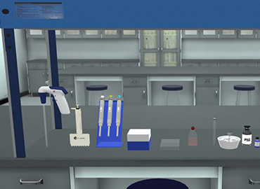
In-Vitro Chromosomal Aberrations Test
Learn how to detect the aberrations in the structural chromosomal by estimating different classes of chromosome changes scored in metaphase.
The used method in this experiment is In-Vitro Screening of Metaphase Chromosomal Aberrations by using Light Microscope.
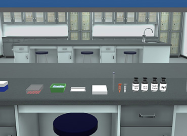
In Vitro Caspase 3 Activity Assay
Understand how to determine apoptotic cells by visualizing the caspase 3 enzyme activity inside cellular nuclei using a fluorescent microscope (caspase-3 activity assay in apoptotic cells). Students will be able to treat the cells with the caspase 3 primary and secondary antibodies, also analyze cells and the resultant data.
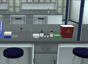
In Vitro Fluorescein Diacetate/Propidium Iodide (FDA/PI) Staining Assay
Learn how to calculate the viability percent of cultured adherent cells after excitation of free fluorescein cleaved by enzymes of esterase in live cells.
By the end of the experiment, students will be able to:
- Understand how to treat cells with the FDA/PI working solution in a cell culture medium.
- Count cells with bright red fluorescence (for dead cells) and bright green fluorescence (for viable cells) by using a fluorescence microscope.
- Understand the concept, principle, and protocol of FDA/PI staining assay experiment.
- Calculate the percent of the viability of viable and dead cells.
- Represent the resulting data of the experiment graphically.
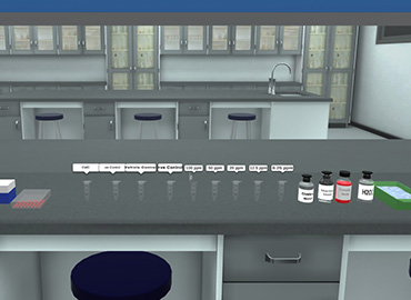
In Vitro Neutral Red Uptake Assay
Learn how to quantify the amount of absorbed supravital dye Neutral Red (NR) in lysosomes of viable cells by using a microplate spectrophotometer (Microplate Reader).
By the end of Neutral Red Uptake Assay experiment, students will be able to:
- Treat the cells with the Neutral Red-cell culture medium.
- Understand the principle, protocol, and procedure of the experiment.
- De-stain the Neutral Red and prepare the extracted solution for analysis.
- Analyze de-stain extracts’ color intensity by a microplate reader and also analyze the resulting data.
- Represent and locate the IC50 of the tested nanoparticles graphically.
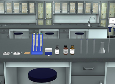
In Vitro Acid Phosphatase Assay for Cell Viability
Understand how to quantify the acid phosphatase activity amount on the cell membrane of viable cells by using a microplate reader.
By the end of the experiment, students will be able to:
- Treat the cells with the solution of the acid phosphatase assay.
- Stop the reaction and measure the color developing calorimetrically.
- Understand the concept and steps of the in vitro acid phosphatase assay.
- Analyze the color intensity of the reaction and the resulting data.
- Represent and locate the IC50 of the tested nanoparticles graphically.
PraxiLabs, the 3D virtual lab solution, provides students with access to realistic biology, chemistry, and physics labs and enriches their understanding with a variety of informational and educational content.
 PraxiLabs A virtual world of science

