Last Updated on March 24, 2026 by Muhamed Elmesery
In today’s evolving medical and science education landscape, hands-on experience no longer has to be confined to a physical laboratory. A fully interactive virtual microscope lab brings the power of real microscopy into a dynamic digital environment—where students can zoom, focus, rotate slides, and analyze specimens with precision and ease.
In this blog post, we will discover what the virtual microscope lab is, its different uses, the subjects that benefit most from virtual microscopy, how the virtual microscope provides a realistic image, the features of PraxiLabs virtual microscope lab, and more!
Table of Contents
What is the virtual microscope lab?
A virtual microscope lab is an interactive and immersive virtual lab that simulates the experience of using a real laboratory microscope—allowing students to observe, zoom, focus, and analyze specimens online.
A microscope lab virtual simulation is a safe, cost-effective, and accessible digital alternative to traditional microscopy, designed to enhance hands-on learning through interactive simulation.
Different uses of the virtual microscope lab:
The main use and benefit of the virtual microscope lab is that instead of physically placing slides under a microscope, learners access high-resolution, magnifiable samples through a computer or tablet.
Users can adjust magnification levels, change focus, switch between lenses, and sometimes even apply staining techniques—just like in a real lab setting.
What subjects benefit most from virtual microscopy?
Medical and Health Sciences
- Histology (Microscopic Anatomy): A core application of online microscope lab, allowing students to study tissues and cells in detail without physical slides.
- Pathology: Used to teach disease processes through digital histopathology slides; students can compare normal and abnormal tissue.
- Cytology: Helps in learning cell morphology and diagnosis; studies show virtual microscopy can be more effective than traditional microscopy for teaching cytology.
- Oral and Dental Medicine: Supports courses in oral histology and oral pathology, improving diagnostic learning.
Biological Sciences
- General Biology: Microscope virtual lab enhances understanding of cellular structure, microbes, and biological specimens under the microscope.
- Parasitology: Useful for identifying and studying microscopic parasites.
Natural Sciences & Laboratory Courses
- Anatomy: Especially microscopic anatomy, where detailed visualization of tissues is needed.
- Hematology: The study of blood cells and their components, benefiting from high-resolution digital slides.
How does the virtual microscope give a realistic image?
The 3d virtual microscope for students is a software system that provides a realistic digital emulation of a high-power light microscope. The raw data for such a system can be captured by digitally scanning collections of full microscope slides under high magnification.
For example, a portion of a digitally captured composite slide, stitched together by hand from multiple individually captured images. While the hardware for performing the data capture more effectively is rapidly becoming commercially available, the software support required to provide interactive response times for the standard behavior of a physical microscope has not been developed.
These behaviors include continuously moving the stage and changing magnification and focus. In addition, a software solution can enable new modes of behavior that cannot be achieved with a physical microscope, such as simultaneous viewing and manipulation of a single slide by multiple users.
Source: https://www.researchgate.net/publication/13871246_The_Virtual_Microscope
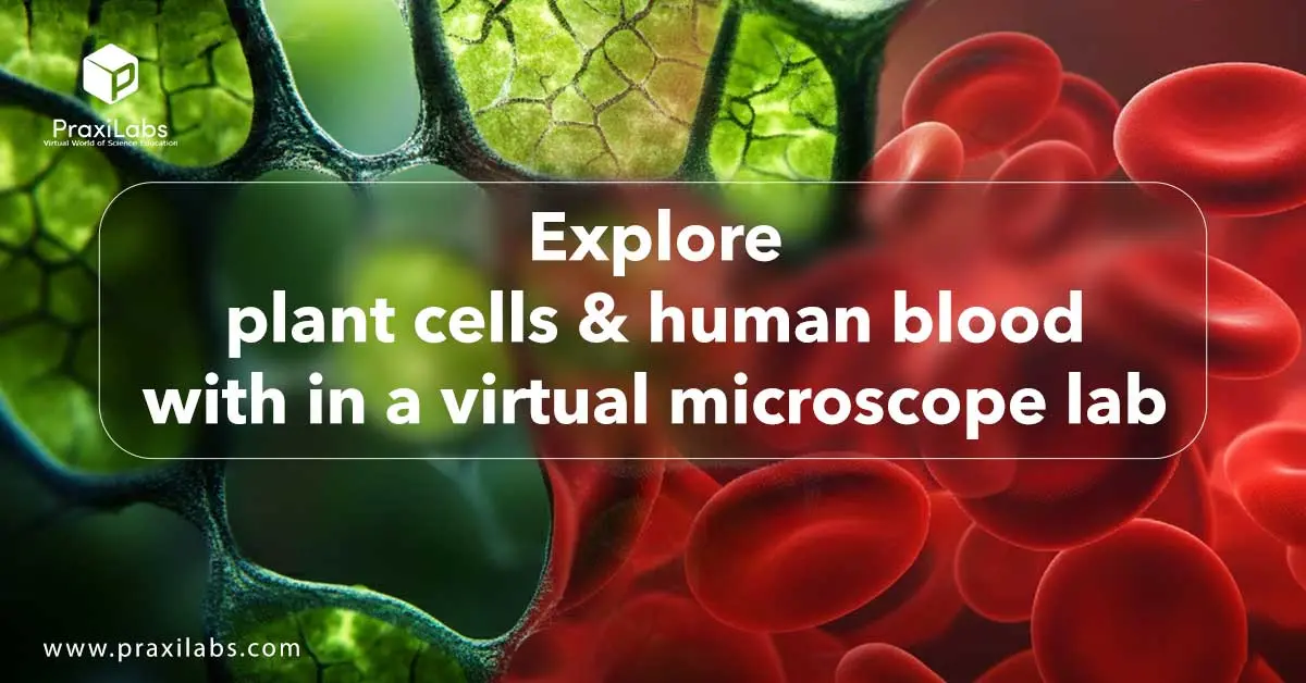
Explore plant cells & human blood within a virtual microscope lab:
With a microscope simulation lab, students don’t just see plant cells and human blood—they actively explore them, zoom into fine details, and connect structure to function in a dynamic, engaging way.
Plant Cells with a Virtual Microscope
Using prepared digital slides, students can:
- Zoom in on cell walls, chloroplasts, nuclei, and vacuoles.
- Adjust magnification levels (4x, 10x, 40x, 100x).
- Compare different plant tissues such as epidermal cells and leaf cross-sections.
- Identify structural differences between plant and animal cells.
This is especially valuable in Botany, Cell Biology, General Biology, and Plant Physiology.
The clear visualization of chloroplast distribution and rigid cell walls strengthens understanding of photosynthesis and plant structure-function relationships.
Human Blood Analysis with a Virtual Microscope
In a digital blood smear slide, students can:
- Differentiate between red blood cells (RBCs), white blood cells (WBCs), and platelets.
- Identify WBC types such as lymphocytes and neutrophils.
- Examine abnormal cell morphology.
- Practice diagnostic observation skills.
This application is widely used in Hematology, Medical Laboratory Sciences, Nursing, Medicine, and Pathology.
Students can repeatedly analyze slides without damaging samples, improving accuracy and confidence in microscopic interpretation.
Features of PraxiLabs Virtual Microscope lab:
In platforms like PraxiLabs, students can explore realistic virtual biology labs where they manipulate microscopes, examine specimens at different magnifications, and conduct complete experiments in a safe, immersive environment.
These virtual simulations from PraxiLabs allow users to:
- Access high-quality slides anytime, anywhere.
- Repeat observations without time limitations.
- Safely explore delicate or rare specimens.
- Strengthen analytical and diagnostic skills.
- Reduce the need for physical microscopes, slides, stains, and consumables.
- Get detailed performance tracking and analytical reports.
- Have hints, explanations, and corrective feedback in real time — especially helpful during virtual microscope activity.
- Create assessment questions related to microscopic observations and lab tasks, with automated grading and feedback.
- Integrate easily with learning management systems (LMS) like Canvas, Blackboard, and Moodle.
Explore the Virtual Biology Lab of PraxiLabs
PraxiLabs’ virtual microscope lab for medical schools & colleges:
Medical schools and college science programs use PraxiLabs’ virtual microscope labs to give students hands-on, repeatable experience with slides and microscopic structures — from human blood and tissues to microbes — all within a safe, curriculum-aligned online environment.
At institutions like Texas A&M University (USA) and Kwantlen Polytechnic University (Canada), PraxiLabs virtual science labs — including microscopy simulations — are integrated into biology courses. Students use virtual microscopes to explore cell structures, tissues, microbes, and other biological specimens, making microscopic observation a core part of remote and hybrid instruction.
With PraxiLabs’ virtual microscope lab, your institution can deliver immersive, repeatable, and curriculum-aligned microscopy training that strengthens diagnostic skills and improves academic outcomes.
- Enhance histology, hematology, microbiology, and pathology courses
- Track performance with detailed analytics and reporting
- Provide 24/7 remote access for hybrid and distance learning
- Reduce equipment costs while maintaining high-quality practical training
Book a demo today and discover how PraxiLabs can elevate your medical curriculum.
Frequently Asked Questions
Can I see bacteria with a virtual microscope?
Yes—high-resolution virtual microscope lab can clearly display bacteria, especially when slides are properly prepared and stained, allowing students to zoom in and observe shape, arrangement, and key structural features.
Are virtual microscopes good for staining differences?
Absolutely. Virtual microscope labs effectively highlight staining variations (such as Gram-positive vs. Gram-negative), making it easier to compare color differences, contrast, and cellular details without slide preparation errors.
Explore Gram Stain Simulation with PraxiLabs