 PraxiLabs A virtual world of science

Last Updated on July 29, 2026 by Muhamed Elmesery
A mitosis and meiosis simulation lab gives students an immersive, hands-on way to explore the mechanisms of cell division without the limitations of a physical laboratory. Understanding how cells replicate and divide is foundational to biology, genetics, and medicine yet these processes are invisible to the naked eye. Virtual simulation labs bridge this gap by bringing microscopic events to life through interactive 3D models, allowing students to observe, repeat, and fully grasp the stages of cell division at their own pace.
Table of Contents
What is mitosis and meiosis simulation lab?
A mitosis and meiosis simulation lab is a virtual environment where students can interact with 3D animated models of dividing cells, observe each stage step by step, and repeat experiments as many times as needed. Unlike static textbook diagrams, these simulations:
- Respond to student input in real time.
- Display phase transitions as they happen.
- Allow zoom-in exploration of chromosomal behavior.
- Transform abstract concepts into concrete, reproducible learning experiences.
Mitosis: Replication for Growth
Mitosis is the process by which a single parent cell divides into two genetically identical daughter cells. It is essential for growth, tissue repair, and asexual reproduction. The lab guides students through all five mitotic stages prophase, metaphase, anaphase, telophase, and cytokinesis with animated chromosome movement at each step.
Meiosis: Division for Reproduction
Meiosis produces four genetically unique haploid cells from one diploid parent cell. It is the foundation of sexual reproduction and genetic variation. Simulation labs display both meiosis I and meiosis II divisions, clearly showing chromosome pairing, crossing over, and independent assortment concepts that are notoriously difficult to visualize in a classroom.
Mitosis and meiosis simulation lab educational aspects:
Virtual labs do not simply replicate physical experiments, they expand what is pedagogically possible. The educational value of these virtual labs spans multiple learning dimensions:
- Visual learning at molecular scale simulations render spindle fibers, centromeres, and chromatids with scientific accuracy. Students rotate, pause, and replay each division phase, dramatically improving retention of complex processes.
- Safe repetition and exploration students re-run experiments, test hypotheses about chromosomal behavior, and explore outcomes without consuming materials or time.
- Immediate feedback and assessment quizzes linked directly to simulations with automated grading highlight which stages need additional reinforcement.
- Accessible bilingual learning experiments available in both Arabic and English, ensuring language is never a barrier to scientific understanding.
How do simulation labs improve genetics education?
Genetics education has historically struggled with one core problem: the phenomena being studied are invisible. Virtual simulation labs directly address this challenge by animating chromosome behavior, connecting genotype to phenotype, and allowing students to model chromosomal abnormalities interactively.
According to Frontiers in Education, active inquiry-based learning the model virtual labs inherently support significantly improves conceptual retention and critical thinking in STEM subjects. Students who engage with simulation-based material outperform peers taught through lecture-only methods.
- Connecting Genotype to Phenotype Simulations animate the relationship between chromosome behavior during meiosis and the genetic traits that appear in offspring.
- Exploring Chromosomal Mutations Students model abnormalities like nondisjunction interactively, building far deeper understanding than textbooks alone.
According to Technavio’s Simulation Learning Market in Higher Education report, the sector is projected to grow by USD 2.32 billion at a CAGR of 20.5% between 2024 and 2029, driven by institutional demand for virtual labs and scalable digital science education.
Benefits of using virtual lab for cell division study:
Institutions that integrate a mitosis and meiosis simulation lab into their biology curriculum gain advantages consistently reported across higher education and secondary science programs:
- Up to 60% cost reduction vs. maintaining physical laboratory equipment, reagents, and consumables
- Up to 80% improvement in learning retention through active interaction rather than passive observation
- 24/7 access to experiments, enabling self-paced study outside scheduled lab hours
- Scalable deployment across campuses and remote learning environments without additional infrastructure
- Seamless LMS integration in as little as 24 hours, minimizing IT complexity
- Real-time personalized guidance via AI lab assistant Oxi
- Elimination of safety risks, making lab access equitable for all students
As documented by Wiley’s learning technology platform, digital interactive tools produce measurable improvements in engagement and academic performance when embedded meaningfully within course design exactly the model that virtual cell division labs support.
Features of PraxiLabs mitosis and meiosis simulation lab:
PraxiLabs offers a dedicated mitosis and meiosis simulation lab within its catalog of +210 science simulations across biology, physics, and chemistry, built for higher education institutions:
- 3D interactive cell division models each simulation renders chromosomes, nuclear envelopes, and spindle apparatus in three dimensions, allowing students to rotate and zoom through every phase of division.
- AI lab assistant: Oxi during any mitosis and meiosis chromosome simulation lab session, Oxi provides real-time personalized guidance, answers student questions, and adapts explanations based on individual progress.
- Custom quiz builder quizzes linked directly to experiments with automated grading and performance tracking.
- Bilingual access all experiments available in both Arabic and English.
- LMS integration Moodle, Canvas, and Blackboard embedded via the cell division simulation lab mitosis and meiosis within 24 hours with minimal technical overhead.
- Global Recognition 11 global awards, 2.5+ million active users, 160+ countries.
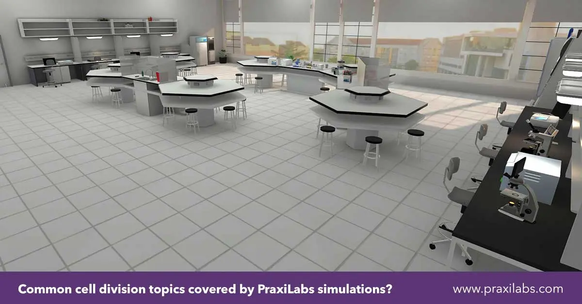
Common cell division topics covered by PraxiLabs simulations:
PraxiLabs covers a curated set of core cell division topics drawn from the biology and genetics curriculum. Below are five key simulations accessible through the PraxiLabs 3D science simulations catalog:
- Mitosis in onion root tip students observe and identify each mitotic phase in plant cells, a curriculum-central experiment brought to life in 3D.
- Meiosis and genetic variation interactive walkthrough of meiosis I and II, showing crossing over and independent assortment with emphasis on genetic diversity.
- Cell Cycle Regulation explores checkpoints controlling cell division and how disruptions lead to abnormal cell behavior.
- Chromosomal structure and organization detailed look at chromatids, centromeres, and telomeres before and during division.
- Nondisjunction and chromosomal abnormalities students model errors in chromosome separation during meiosis, connecting molecular events to real-world genetic conditions.
Explore Onion Root Tip Mitosis Virtual Lab with PraxiLabs
According to Verified Market Research on virtual laboratory platforms, the global virtual and remote laboratories market is expanding rapidly, driven by institutional demand for scalable digital science education tools that do not compromise on experimental rigor.
Frequently Asked Questions
What are the main types of cell division?
The two main types are mitosis and meiosis. Mitosis produces two genetically identical daughter cells for growth and tissue repair. Meiosis produces four genetically unique haploid cells for sexual reproduction. A mitosis and meiosis simulation lab lets students explore both processes from the cell division simulation lab mitosis and meiosis perspective through detailed 3D animations of each division stage.
How to do a mitosis experiment?
In a mitosis and meiosis simulation lab, students prepare specimens, observe cell phases through a 3D microscope interface, and record findings without physical materials, staining chemicals, or microscope equipment.
How is mitosis different from meiosis?
Mitosis produces two diploid identical daughter cells in somatic cells for growth and repair. Meiosis produces four haploid unique cells in reproductive cells via crossing over and independent assortment. A mitosis and meiosis chromosome simulation lab makes these distinctions visually explicit, allowing students to compare chromosomal behavior across both processes side by side.